Quality of the image representation of stent structure, stent enhancement algorithm, in assessing the optimal prevalence of stent in long chronic lesions
DOI:
https://doi.org/10.48026/issn.26373297.2025.1.16.1Keywords:
stent enhancment, invasiv cardiology, PCI, coronary stent, coronary stent restenosisAbstract
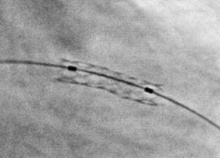
During percutaneous coronary intervention, proper stent implantation is often challenging. Stent enhancement (SE) may be used to visualize implanted stents. SE is an angiographic technique that digitally enhances the visibility of stent metal struts, in order to better assess whether it has been adequately implanted. It is a faster and cheaper alternative to intravascular methods (IVUS, OCT), but it is still less detailed. We perform SE after stent implantation using balloon markers; and it can help us to verify some complications or abnormalities after stent implantation such as stent malposition (stent not pressed against vessel wall), insufficient stent expansion (the stent not being sufficiently expanded), stent deformation (mechanical change of stent shape), edge dissection at stent edge, or stent fracture (stent-structure break). Any of these complications significantly increase the risk of in-stent occlusion or stenosis.
Hypothesis: The change in electrical conditions of diascopy and image acquisition significantly affects the quality of the SE representation of the stent structure
Patients: The study was conducted at the Department of Invasive Cardiology, University Clinical Center of Sarajevo (KCUS), from January 1, 2022, to December 31, 2022. The study included patients of both sexes and different age structures. Hemodynamically stable patients were
included in the study, who underwent stenting of chronic lesion on coronary
vessels with stents longer than 18 mm of different diameter.
Methods: The procedures were performed in the cardiac catheterization laboratory using a TOSHIBA INFINIX fluoroscopic system. The stent is imaged natively, without contrast application, for 4 seconds to acquire a minimum of 34 images. After deflation, the stent balloon must not be moved until the native imaging is completed, as the reconstruction software performs image stacking by marking the balloon markers.
Results: In the control group of 287 participants, selected based on the study inclusion criteria, the image quality was evaluated. There were 196 (68.6%) males and 91 (31.4%) females. By applying the chi-squared test, a statistically significant difference was found in the gender distribution of participants, with males being more prevalent, χ² = 7.078; p = 0.008. According to the subjective assessment of image quality with implanted stents, the evaluators rated the images on a scale from 1 to 5. The quality scores of two groups of images (Table 3) were compared: with (90 kV and 900 mA) and (150 kV and 600 mA), where a statistically significant difference was found in the image quality score of the two groups depending on the electrical potential (p<0.05).
Conclusion: The stent strut visualization using the SE algorithm can be useful in assessing proper stent implantation and determining the potential need for post-dilatation. The use of the SE algorithm is limited due to artifacts caused by pacemaker electrodes or patient obesity, as well as cardiac motion artifacts that prevent SE reconstruction in certain positions. According to the study results, stent strut visualization is of better quality at higher mA values and lower kv values.
Downloads
Published
How to Cite
Issue
Section
License
Copyright (c) 2025 Haris Porobić, Mirad Hujdur; Darko Tomić; Sabina Prevljak, Armin Papračanin, Merjema Imamović, Nihad Kukavica

This work is licensed under a Creative Commons Attribution 4.0 International License.
Copyright & licensing:
This journal provides immediate open access to its content under the Creative Commons CC BY 4.0 license. Authors who publish with this journal retain all copyrights and agree to the terms of the above-mentioned CC license.
