Comparison of mammography and ultrasound in preoperative assessment of suspected breast lesion
DOI:
https://doi.org/10.48026/issn.26373297.2025.1.16.2Keywords:
mammography, ultrasound, histopatology, breast cancer, tumor sizeAbstract
Aim of the study: The aim of this study is to present the role of mammography in assessing the size of malignant breast lesions, to present the role of ultrasound in the evaluation of malignant breast lesions, and to demonstrate mammography as the gold standard in the assessment of breast cancer. A retrospective-prospective study was conducted at the Radiology Clinic of the Clinical Center of the University of Sarajevo. The study included 50 female patients with a mean age of 52.94 ± 11.46 years, the youngest patient was 32 and the oldest was 77 years.
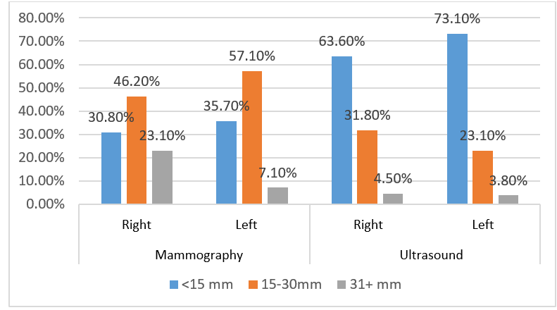
Results: Analysis of the reasons for visiting a doctor showed that 21 patients (42%) had no prior diagnosis, while 24 patients (48%) came for follow-up examinations. All patients underwent mammography imaging, breast ultrasound examination, and histopathological (HP) analysis. The analysis revealed that, out of 40 evaluated mammographic images of the right breast, a suspicious lesion was identified in 27.5% of the images. Analysis of the distribution of patients according to the BI-RADS score showed that BI-RADS categories 1 and 2 were observed in 48.89% of cases on ultrasound and in 55.32% of cases on mammography. BI-RADS categories 5 or 6 were observed in 6.66% of patients on ultrasound and in 8.59% of patients on mammography. There was no significant difference in the distribution of patients (x2=3.214, p=0.782). Based on the available data on the size of histopathological samples, it was determined that 40.91% were smaller than 15 mm, as well as within the range of 15 to 30 mm. Histopathological samples larger than 30 mm accounted for 18.18% of the cases. The analysis showed that, out of 50 histopathologically examined samples, 35 (70%) had a benign diagnosis, 6 (12%) patients had a suspicious lesion, and 9 (18%) patients were diagnosed with carcinoma. The analysis also revealed that the sensitivity of ultrasound was 44.44%, with a confidence interval of 13.70 to 78.8%, while the sensitivity of mammography was 55.56% (21.20–86.30%). The specificity of ultrasound was 97.14%, with a confidence interval of 85.08–99.93%. Specificity of mammography was 97.37% (86.19-99.93%). The positive predictive value was 80% for ultrasound and 83.33% for mammography.
Downloads
Published
How to Cite
Issue
Section
License
Copyright (c) 2025 Zerina Pojskić

This work is licensed under a Creative Commons Attribution 4.0 International License.
Copyright & licensing:
This journal provides immediate open access to its content under the Creative Commons CC BY 4.0 license. Authors who publish with this journal retain all copyrights and agree to the terms of the above-mentioned CC license.
