Savremene dijagnostičke metode u nuklearnoj medicini - detekcija CNS oboljenja PET metodom
Keywords:
PET-CT, CNS, nuclear medicineAbstract
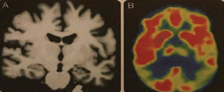
Nowadays PET is intensively used in the research of different disorders of central nervous system (CNS). A whole series of the positron radiopharmaceuticals has been developed for the noninvasive examination of the brain metabolism of glucose, cerebral blood flow, neurotransmitter systems, usually labeled as 11C, 13N, 18F. The positron emission tomography (PET) is an important method in the functional imaging of the brain that enables in vivo researches of brain functions, and it is possible to research almost every aspect of brain functions due to present-day development. In clinical practice, but in researches, as well, the most used is 18F-fluoro-deoxyglucose (FDG). Physical characteristics
enable easier production and usage, and it is used for studying and estimation of disorders of glucose metabolism in many pathological conditions of the brain.
In the past PET was mostly used in the researches because of relatively high cost and complexity of accompanying infrastructure, such as cyclotrons, PET camera and radiochemical laboratory. In the last few years due to the development in the technology and prevalence of PET cameras, PET is more and more used in clinical neurology enabling the better comprehension of the disease and pathogenesis, it facilitates establishing a diagnosis, it enables prognostics following the development of the disease, as well as the response to the applied therapy. Special advantages of the molecular imaging lie in the fact that sophisticated biological processes and specific ways in certain disease can be explained on cellular and molecular level in human and other living systems. Besides, molecular imaging can provide the information on clinical changes before the pathological factors appear, thus enabling the diagnosis of the disease in the early stages and the help in the therapeutic examinations of many CNS disorders.
Downloads
Published
How to Cite
Issue
Section
License
Copyright (c) 2019 Radiološke tehnologije

This work is licensed under a Creative Commons Attribution 4.0 International License.
Copyright & licensing:
This journal provides immediate open access to its content under the Creative Commons CC BY 4.0 license. Authors who publish with this journal retain all copyrights and agree to the terms of the above-mentioned CC license.
